Green Fluorescent Nissl Stain
| 貨號 | 數量 | 價格 | 交貨時間 | 購買此產品 |
|---|---|---|---|---|
| 1D010 | 50 uL |
–
|
12 天 | |
| 2D010 | 500 uL |
$139.00
|
現貨 |


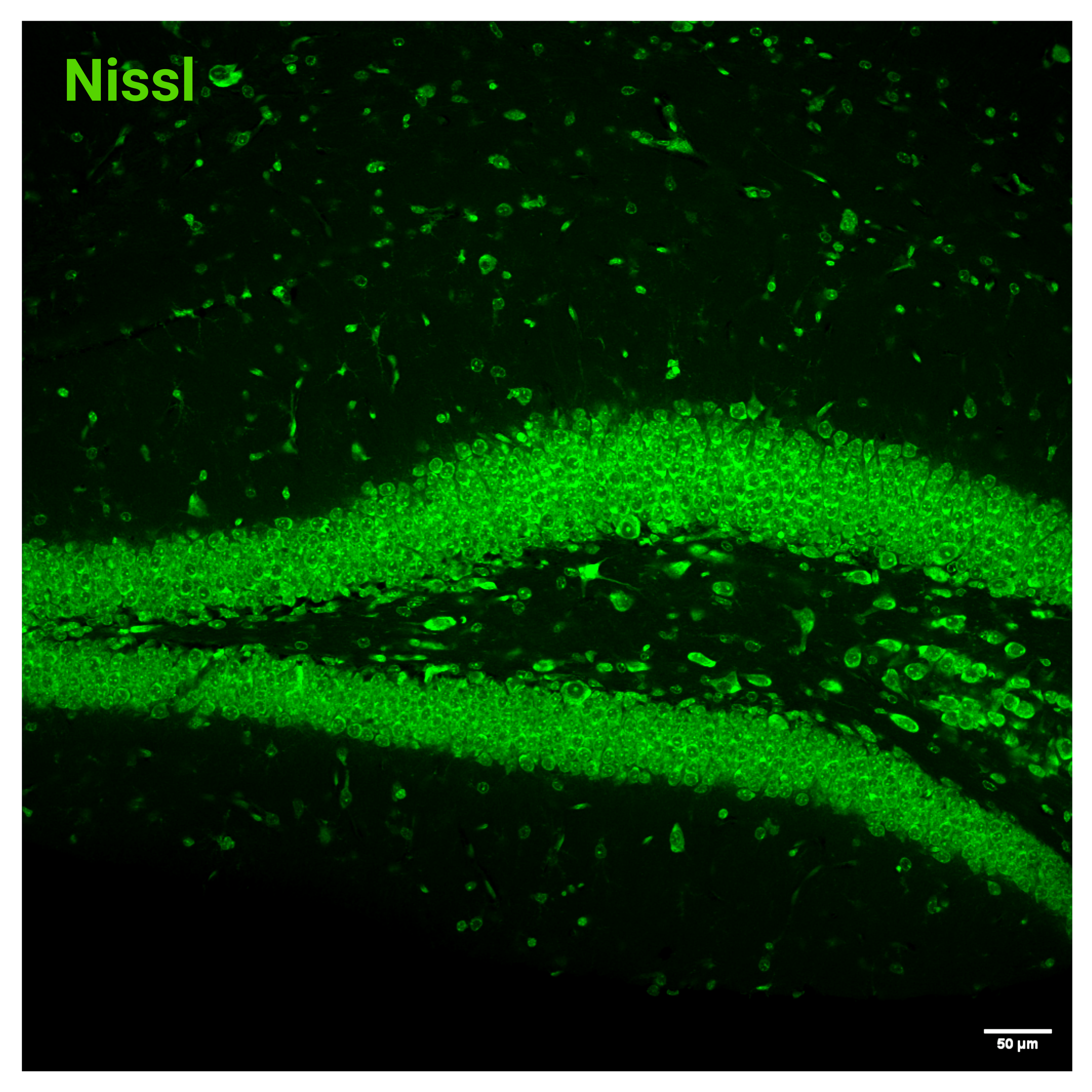
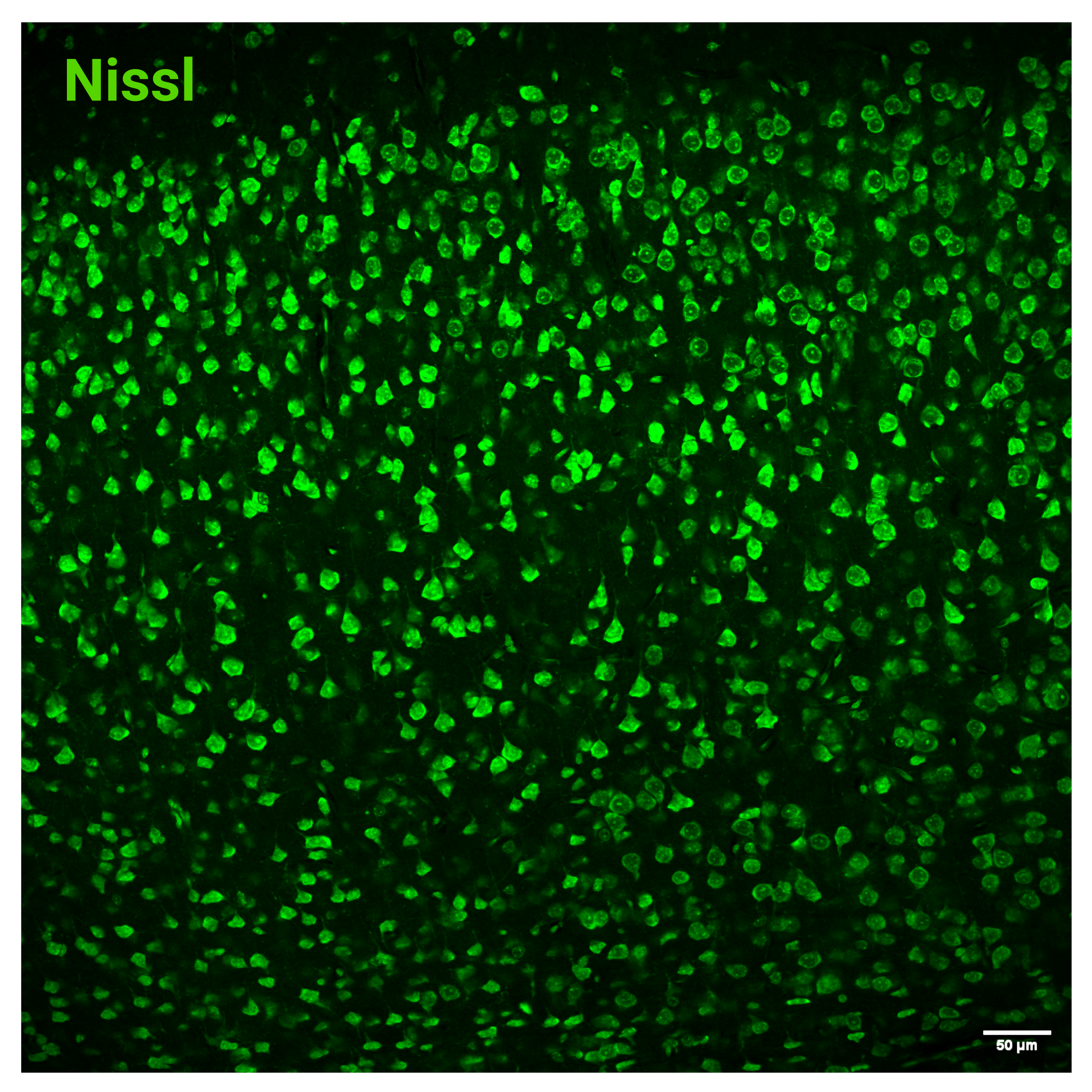
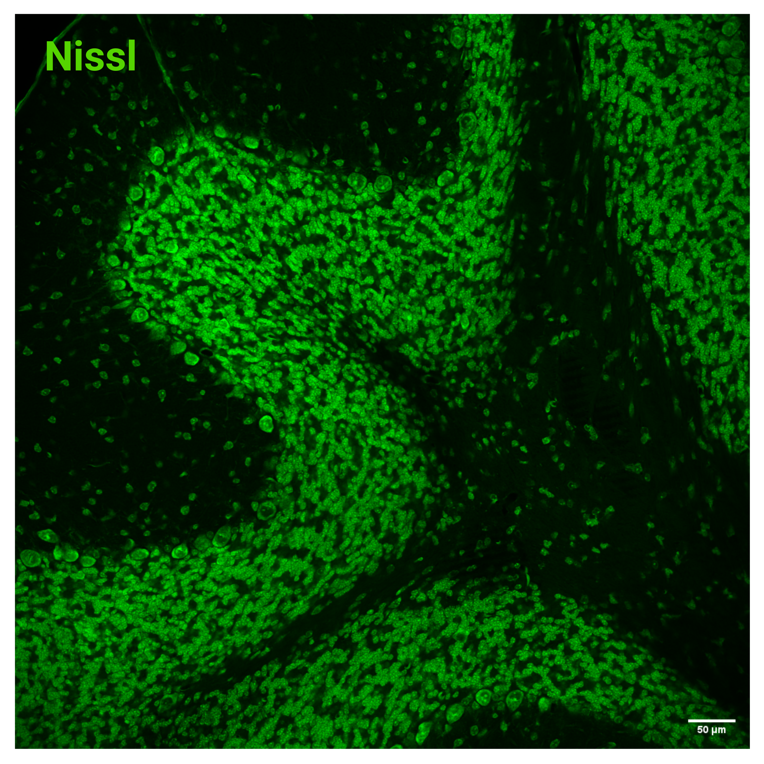
Nissl staining is a widely used histological method for visualizing the morphology and cytoarchitecture of nervous tissue. The technique is based on the selective staining of Nissl substance, a structure rich in ribosomal RNA within the rough endoplasmic reticulum of neurons. As a result, neuronal cell bodies are labeled much more intensely than surrounding cells, facilitating the identification of neuronal populations and the assessment of neural tissue organization.
We offer highly concentrated (1,000×) Fluorescent Nissl Stains with different spectral properties.
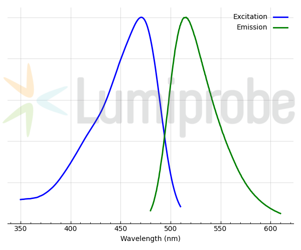
Green Fluorescent Nissl Stain (also known as Fluoro Nissl Green) is a cell-impermeant fluorescent dye that exhibits low background fluorescence in solution and becomes highly fluorescent upon binding to nucleic acids. The dye effectively labels RNA-rich neuronal cell bodies in fixed tissue sections, producing bright green fluorescence with excitation and emission maxima at 471 nm and 515 nm, respectively.
The spectral properties of Green Fluorescent Nissl Stain make it compatible with multicolor fluorescence imaging. Its emission is well separated from blue fluorophores such as DAPI and Hoechst dyes and from orange- and red-emitting probes including AF 594, Cyanine3, Cyanine5, and related fluorophores, allowing flexible integration into complex imaging experiments.

染色標題
顧客還購買了這個產品


General properties
| Appearance: | yellow solution |
| Molecular weight: | 248.29 |
| CAS number: | 161622-27-1 |
| Molecular formula: | C15H12N4 |
| IUPAC name: | 10H-Indolo[2-b]quinoline-3,8-diamine |
| Quality control: | NMR 1H and HPLC-MS (95+%) |
| Storage conditions: | 24 months after receival at -20°C in the dark. Transportation: at room temperature for up to 3 weeks. Desiccate. |
| MSDS: | 下載 |
| 產品規格 |
Spectral properties
| Excitation/absorption maximum, nm: | 471 |
| Emission maximum, nm: | 515 |


 $
$ 
